PLANT CELL AS EASY SYSTEM TO ANALYSE OSMOSIS
Questo articolo scientifico rappresenta il risultato del lavoro dei ragazzi di 4C scientifico di Melzo, che hanno sperimentato cosa significa fare Scienza, tenendo insieme l’aspetto teorico (imprescindibile) e quello pratico/laboratoriale
4C Class school year 2021-22* and Prof. Galliani*
*Liceo linguistico scientifico statale “Giordano Bruno”, Melzo
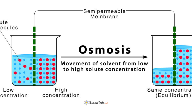
L’osmosi è un movimento di acqua (solvente) da una soluzione meno concentrata (ipotonica) ad una più concentrata (ipertonica), attraverso una membrana semipermeabile, cioè una membrana che permette solo il passaggio del solvente e non dei soluti come la membrana plasmatica. Si tratta di un processo spontaneo, che avviene cioè senza apporto esterno di energia.
L’allestimento di campioni vegetali «a fresco» o in vivo, permette di seguire eventi dinamici come l’osmosi. La cellula vegetale con vacuolo colorato oppure ricca di cloroplasti è un sistema facilitato per l’osservazione dell’osmosi. Infatti, i movimenti di acqua tra interno ed esterno della cellula, che non sono direttamente visibili, possono essere osservati indirettamente in quanto determinano variazioni del volume colorato del vacuolo oppure variazioni del volume del citoplasma e conseguente spostamento dei cloroplasti colorati di verde.
RESULTS
In a hypotonic solution, with lower magnification (4X), the thin layer of the radicchio rosso epidermis appears formed by a homogeneous red layer (figure 8a). Then, with higher magnifications (10 X and 40 X), turgid red cells, tightly linked to each other, become evident (figure 8b and 8c). The boundaries of the cells appear white and are clearly visible thanks the presence of the cell wall and of the cytoplasm that is localized only at the periphery of the inner environment because of the high turgor pressure (figure 8c). Almost all the inner environment is occupied by the coloured vacuole.

Finally, at higher magnification (40 X) it is also possible to see some characteristic structures of a plant cell:
- the nuclei of the cells appear like a small round and translucent objects in the inner environment;
- the trichomes (leaves “hair”) are fine outgrowths formed by thin cells that can have different specialized roles like the defence from herbivores, the reduction of transpiration and the reflection of sunlight protecting the more delicate tissues;
- the stoma is a pore in the epidermis of leaves, which controls the rate of gas exchange. The pore is bordered by a pair of specialized parenchima cells known as guard cells that are responsible for regulating the size of the stomatal opening
In a hypertonic solution, with lower magnification (4X), the thin layer of the radicchio rosso epidermis appears formed by a heterogeneous layer, all white with many red dots.
Then, with higher magnifications (10 X and 40 X) the cell wall that surrounds each cell like a thin black boundary becomes visible. The red spots concentrated in the inner environment are the restricted vacuoles that have lost water. The white region surrounding the vacuole is occupied by the shrunk cytoplasm and by the plasma membrane detached from the cell wall. This process is called plasmolysis and that is why plants lose turgor pressure and wilt. In some cases, it is possible to recover it by giving hypotonic solution to the plant tissue.


Osmosis process can be also observed in a living leaf of the aquatic plant Elodea canadensis.
Very thin leaves characterize this plant and make it a suitable plant organism for direct microscope analysis without any preparation. Therefore, it is possible to observe chloroplasts, the organelles characterized by two membranes and a high concentration of chlorophyll, a green pigment essential to conduct photosynthesis.
Under the microscope, a phenomenon called cytoplasmic streaming, in which cytoplasm and organelles such as chloroplasts move throughout the cell, can be monitored. This process changes visibly when the cells are immersed in solutions with different concentration. In a hypotonic solution, the inner environment of plant cells expands until they become turgid for the presence of cell wall. As consequence, green chloroplasts are equally diffused in all the inner environment (figure 11a). On the other hand, in hypertonic environment the plant cells lose water and shrink. Consequently, the vacuole (that is not visible) decreases in size and the plasma membrane detaches from the cell wall causing the citoplasma to constrict. This process is made visible under light microscope because the green chloroplasts are concentrated in a small region of the inner environment (delimitated by cell wall).

DISCUSSION AND CONCLUSIONS
The preparation of “fresh” or in vivo plant samples allows to follow dynamic events such as osmosis. This analysis is not possible with animal tissues because animal cells don’t have cell wall that gives them rigidity and keeps them attached. Moreover, animal cells don’t have structures already coloured that make easy the observation with light microscope.
The vacuole, present only in plant cells, is the organelle involved in the regulation of water movements between the inside and the outside of the cell. The plant cell can change the concentration of the vacuole solution and so influence the osmotic movement of the water between the inside and the outside of the cell. Furthermore, the vacuole can contain coloured pigments, which give colour to petals or fruits.
In this study, we found that plant cells are an easy system to observe the phenomenon of osmosis. The water movements between the inside and the outside of the cell can be observed indirectly under the light microscope by exploiting the cells that contain pigments in the vacuole. Actually, the transit of water determines variations in the volume of the coloured vacuole.
Osmosis process can be also observed in a living leaf of the aquatic plant Elodea canadensis. Under the microscope, a phenomenon called cytoplasmic streaming, in which cytoplasm and organelles move throughout the cell, can be monitored in different external conditions directly following the movement of green chloroplasts.
In addition to this, simple experiments that use common and cheap objects, as the ones we carried out, allows students to learn complex scientific concepts more easily and smartly.
AKNOWLEDGMENTS
We thank Ombretta Breno, the lab technician of the GB school in Melzo, for her essential support for the preparation of materials and for the realization of the didactic activity. We also thank Prof.ssa Zamarioli for the English revision of the manuscript. This study was funded by Liceo statale linguistico scientifico “Giordano Bruno”. Approved by GB EXPRESS as journal paper n° 4.